

Overview
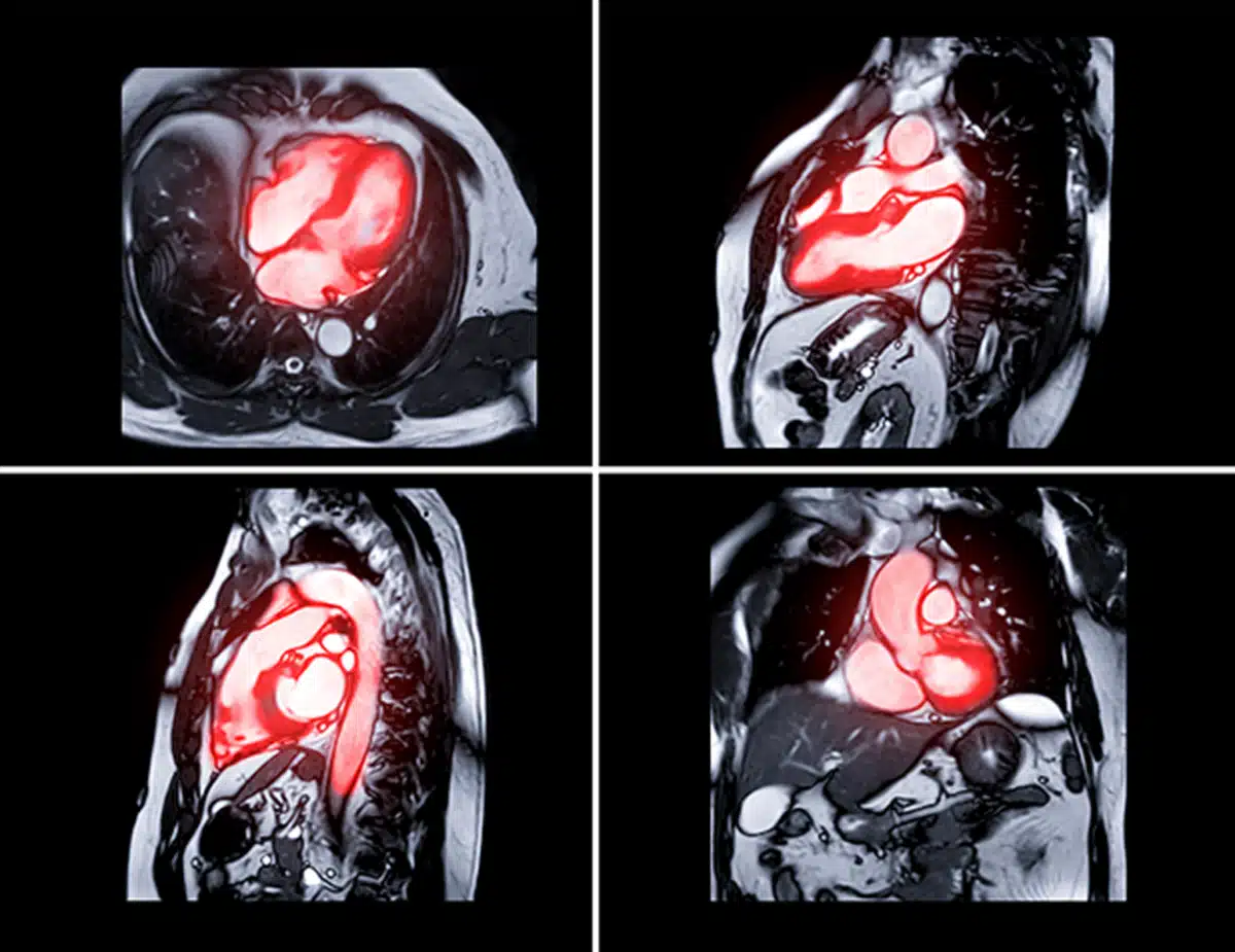
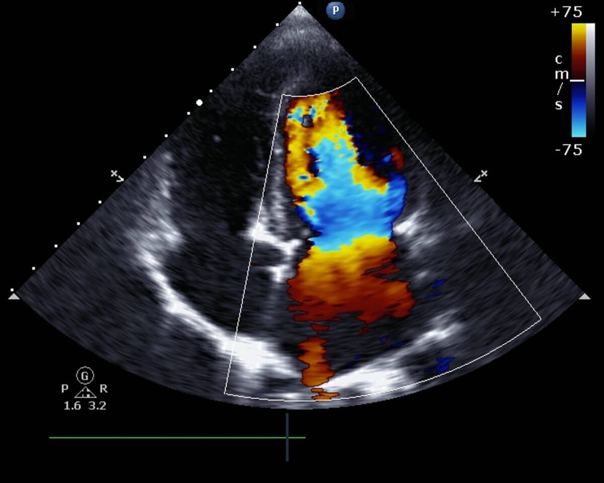
Clario’s cardiovascular imaging team has experience with all imaging modalities including the latest innovations in 3D and molecular imaging, cardiac MRI, echocardiography, and myocardial strain to support evaluation of your drug or device for sensitive detection of both efficacy and safety signals.
Cardiovascular expertise
You begin an ongoing relationship with our multidisciplinary team when you partner with Clario for cardiovascular imaging support. We include scientific experts to ensure your imaging plan meets your operational milestones yet provides the most robust data. In the planning phase, our client services team works with scientific and regulatory experts to review the study design, and suggest protocol options that maintain your trial’s integrity, while respecting budgetary and timeline concerns.
Clario’s scientific team comprises highly qualified MDs, PhDs, and technologists staff with extensive experience who ensure a high degree of rigor throughout all phases from study design to investigational site training, image acquisition, and final expert central assessment. Board-certified cardiologists, radiologists, nuclear cardiologists, registered radiologic technologists, and sonographers provide expert independent reviews with extensive quality assurance checks.
This integrated approach enables us to:
- Ensure image quality and consistency from the outset with specialists guiding the image protocol development process
- Improve site quality and limit variability, which is a critical factor determining the success or failure of a clinical trial. Clario’s tailored and comprehensive site support maximizes image quality enhancing the interpretability of your trial
- Conduct initial image assessment using modality-specific registered sonographers/technologists trained to ensure precise and reproducible measurements
- Expert physician over-reads to confirm all assessments and ensure endpoint fidelity
- All expert physicians and technologists are certified within a cardiovascular medicine subspecialty for each required modality
- Develop rapid comprehensive data transfers that support the approval process

Expert independent review and quantitative analysis
Our independent reviewers have extensive experience with the latest advances in the evaluation of cardiac structure and function. Clario’s specialists analyze all images from baseline through follow-up and support a wide range of imaging modalities.
Regulatory guidance
Clario’s experienced regulatory team participates in every step of the clinical trial process, providing expert guidance for FDA, EMA, PMDA and CFDA submission.
Robust imaging data
Clario maintains rigorous quality control throughout the trial to ensure high-quality data, including:
- Formal qualification of imaging sites and equipment
- Specification and customization of image acquisition requirements for all modalities
- Imaging protocol training and ongoing technical support and site feedback
- Quality control evaluation for every imaging timepoint
- A unique approach to fiducial point analysis limiting variability and increasing precision for each measurement
Clario supports a wide range of imaging modalities and quantitative cardiovascular imaging endpoints across all phases
300+
Cardiovascular clinical trials
30+
Cardiovascular indications
70+
Non-cardiovascular endpoints
4,000+
Sites supported
Indications and areas utilizing cardiovascular imaging
Our recent work includes imaging support for the following:
Cardiovascular indications
- Abdominal Aortic Aneurysm
- Amyloidosis
- Anemia
- Atrial Fibrillation
- Cardiomyopathy
- Cardiovascular Contrast Agent
- Chemotherapy-induced Cardiotoxicity
- Danon Disease
- Giant Cell Arteritis
- Heart Disease
- Heart Failure
- Hypertension
- Idiopathic Pericarditis
- Left Ventricular Hypertrophy (LVH)
- Left Ventricular Systolic Dysfunction
- Myelodysplastic Syndrome
- Myocardial Infarction (MI)
- Myocardial Perfusion
- Peripheral Arterial Disease (PAD)
- Peripheral Vascular Disease (PVD)
- Pulmonary Arterial Hypertension (PAH)
- Pulmonary Embolism (PE)
- Sickle Cell Disease
- Takayasu Arteritis
- Thromboembolic Disorder
- Thrombosis
- Valvular Regurgitation
Non-cardiac indications utilizing cardiovascular imaging
- Alzheimer’s Disease
- Asthma
- Chronic Kidney Disease
- Chronic Obstructive Pulmonary Disease (COPD)
- Chronic pain
- Diabetes
- Epilepsy and associated syndromes
- Lennox Gastaut Syndrome
- Myloclonic Astatic Epilepsy (Doose Syndrome)
- CDKL5 Deficiency Disorder
- Genetic Cardiomyopathies
- Dravet Syndrome
- Rett Syndrome
- Friedreich’s Ataxia
- Genetic storage diseases
- Mucopolysaccharidoses (MPS) VII
- Fabry
- Acid Sphingomyelinase Deficiency (ASMD)
- Muscular Dystrophy
- Hunter Syndrome
- Pompe Disease
- Methylmalonic Acidemia, Propionic Acidemia
- Huntington’s Disease
- Gaucher’s Disease
- GM2 Gangliosidosis
- Hemodynamic Organ/Systemic Diseases
- Cirrhosis
- Bacterial Infections
- Hepatitis C
- Idiopathic Pulmonary Fibrosis
- Inflammatory Diseases
- Rheumatoid Arthritis
- Ulcerative Colitis
- Migraines
- Obesity
- Oncology
- Rare Bone and Muscle Diseases
- Cirrhosis
- Bacterial Infections
- Hepatitis C
- Idiopathic Pulmonary Fibrosis
- Toxic/Metabolic Diseases
- Primary Hyperoxaluria
- X-linked Hypophosphatemia
Expert cardiovascular modality support
Clario’s experience covers the complete range of imaging modalities for cardiovascular research, including:
- Cardiac Magnetic Resonance Imaging (CMR)
- Cardiac Computed Tomography (CCT)
- Computed Tomography Angiography (CTA)
- Cardiac and Vascular Ultrasound (US)
- Multigated acquisition (MUGA)
- Cardiac Positron Emission Tomography (PET)
- Single Photon Emission Computed Tomography (SPECT)
- Echocardiography (ECHO)
- 2D, M-Mode, and 3D imaging
- Color, pulse wave, and continuous wave doppler
- Comprehensive tissue doppler indices
- Multi-chamber global, segmental, and circumferential strain
State of the art cardiac structure & function analysis
- Systolic and diastolic function assessments (i.e., left/right ventricular ejection fraction, cardiac volumes)
- Valvular and hemodynamic assessment
- Myocardial perfusion (viability, infarct size, scar)
- Coronary artery calcium, angiographic, and flow evaluation (CT/Angiography)
- LV/RV/LA strain analysis (CMR/ECHO)
- Tissue characterization including T1/T2 mapping, extracellular volume, quantitative fibrosis, etc. (CMR)
Meet the team
-

Tonya Varcelli, Ph.D.
Director, Scientific and Medical Affairs
-

Joyce Suhy, Ph.D.
EVP Medical Imaging and Specialty Solutions
-

Wan Chi (Gigi) Lau
VP, Medical Imaging